The Inner Ear (Cochlea) is where transduction takes place.
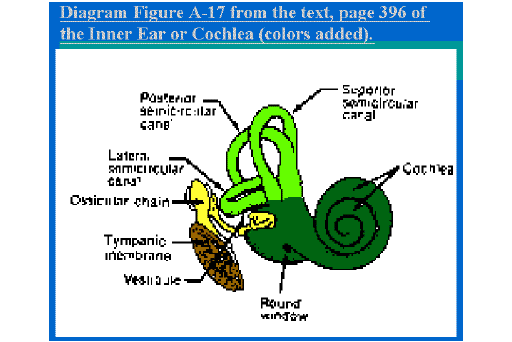
The Inner Ear: The function of the out and Middle ear was to conduct sound energy to the Inner Ear where the actual transduction takes place. The Inner Ear is usually referred to as the Cochlea (in dark green in the picture on the next screen).
It is basically a tube filled with fluid and embedded in the Temporal bone. Unlike the porous bone surrounding the Middle Ear Cavity, the Temporal bone has the highest density of any bone matter in the body.
This is probably a testimony to the delicate nature of this mechanism and its need for protection.
The tube is wound around a central core so that it would resemble a snail shell. At its base are three other tubes, which we have discussed before. These are the Semi Circular Canals of the Vestibular sense (in light green in the picture on the next screen).
Diagram Figure A-17 from the text, page 396 -- the Inner Ear or Cochlea (colors added).
In the grahic view you may see a diagram of the inner ear with the cochlea in dark green.
The Cochlea is a double layered tube filled with fluid.
The Cochlear tube is actually divided in half, length wise. If it is visualized as being straight, a large scale model would resemble a long classroom on the second floor, with a classroom of equal size directly beneath it. At the back of the room (which would be the top of the snail shell) is a stairway leading down into the room beneath. The room beneath is empty except that it is filled with fluid.
Fluid also fills the classroom above.
There is, in addition, at the front of the room below, a round window which is covered with a membrane. This serves as a pressure release when sound waves enter the fluid in the room.
At the front of the room on the second floor, is an oval window covered by a membrane through which the third ossicle (the Stapes) punches in its vibrations.
The floor of the Cochlea has the Basilar Membrane on which are four rows of hair cells.
The vibrations, generated by the third ossicle, set up waves in the fluid which proceed to the back of the room (top of the snail shell), down the stairs, and to the front oval window of the room below.
The floor that separates these rooms is hard (bone tissue) except for a long strip that runs lengthwise up the side. The consistency of this strip is reminiscent of a trampoline. It is called the Basilar Membrane.
In the top classroom, there are four rows a chairs that occupy the area we are calling the Basilar Membrane. If you imagine students sitting in these chairs (with scuba gear, of course), these would be analogous to the four rows of hair cells in the Cochlea.
In our model there might be space for each row to be 40 chairs deep each. In the Cochlea, however, the depth would number in the thousands.
The celia on some hair-cells on the Basilar Membrane are bent by each sound wave that enters the fluid.
There are other cells in the Cochlea, but their function is to support the hair-cells and the transduction process. It's a pretty nice life for a hair-cell except for one inconvenience. Their celia (hairs) are embedded in another membrane called the Tectorial Membrane.
In our classroom model, this would be analogous to a large role of fly-paper stretched from the front to the back of the room across the heads of everyone sitting on the Basilar Membrane.
Hence, when a sound wave is transmitted into the room by the Stapes, the cells on the Basilar membrane rock back and forth like tourists on a raft.
BUT...because their hairs are embedded in the fly paper (Tectoral Membrane) the hair cells receive a wrenching with each wave movement.
Because of the structure of the air cells and the composition of the fluid around them, each wrenching generates an electrical potential in the hair-cell.
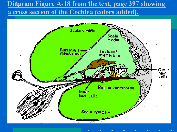
Diagram Figure A-18 from the text, page 397 -- a cross section of the Cochlea (colors added).
In this diagram you can seof the Basilar Membrane In dark brown,and the Hair Cells in yellow and the Tectorial Membrane in light brown.
The hair-cells along the Basilar membrane respond selectively to specific frequencies.
The physical wrenching of the hair-cells, and the corresponding generation of an electrical potential is the actual instance of transduction.
Not all of the hair cells, however, are stimulated by a sound wave. Two factors account for this. One is that the Basilar membrane is actually thicker (more mass) towards the back of the room (top of the snail shell). Hence, it resonates more for low frequency vibrations.
The Base of the Cochlea resonates more for high frequency sounds. The other is that low frequency sound waves tend to be longer and more penetrating, whereas high frequency waves are short.
The result is that the hair-cells along the Basilar membrane perform much like the keys to a piano. That is, hair-cells at specific locations respond to specific frequencies.
A hair-cell and the nerve connected to it is called a Basic Sensory Unit.
A hair-cell and the nerve that connects with it is called the Basic Sensory Unit. If either of these is damaged, a sensory-neural hearing loss will result.
This is quite a different kind of loss than the conductive loss which we discussed earlier. Here are three differences.
1) The first, is that although a sensory neural loss may be so mild that people do not even know that they have it, it can also, unlike a conductive loss, be total, with no residual hearing remaining at all. Granted this is rare, and there is usually some residual hearing. However, this often goes undeveloped and unused by deaf individuals.
2) The second, is that unlike a conductive loss which is generally equal across all frequencies, a sensory neural loss typically effects some frequencies more than others. And almost always, it is the higher frequencies that are typically effected first.
A sensory neural loss usually begins in the higher frequencies and works it way down.
A significant high frequency loss of 6000 Hz and higher would probably not be noticed by a person even if it occurred in both ears. If would be of medical significance, however, because it is a "red flag" heralding a probable progressive hearing loss.
A doctor would want to explore the possible agents that might be contributing to the loss; to consider medication or changes that might be made in the environment to stop the progression of the loss; to set up a schedule of re-tests to monitor any changes in the loss that occur over time; and to prepare individuals how to cope with a hearing loss.
As the loss dips down into the speech range, the individual progressively loses the ability to hear consonants. For young children developing language, this is particularly insidious. Because they can hear the lower frequencies very well, they respond to sounds...even soft ones. Hence, no one ever guesses they have a major hearing loss.
An Audiogram is a chart of hearing ability.
A profile of hearing ability is recorded on a chart called an Audiogram. Typically, an audiogram has numbers across the top which show each frequency (pure tone) that will be sampled. For example they might include: 125, 250, 500, 1000, 2000, 4000 and 8000 Hz.
On the left, going down the side, would be the intensity, marked off in decibels relative to threshold, required for a person to hear each pure tone at threshold.
These might include: -10, 0, 10, 20, 30, 40, 50, 60, 70, 80, 90, and 100 dB.
A series of pure tone are presented with increasing intensity. The individual raises his hand when he finally hears it. Most audiograms use an "X" to she the threshold for the right ear and an "O" for the left ear.
If one connects the points with a line, a profile of hearing is apparent. In the following audiograms you can see the high frequency loss in the profile.
An audiogram showing a significant loss above 4000 Hz.
See an audiogram showing a 4000Hz loss and above. Speech is still intelligible but many fricatives are not heard.
Amplification will not always improve speech discrimination affected by a sensory neural loss.
There are many causes of sensory neural loss in infants an children.
This includes viral infections of the mother before birth or the baby after; blood incompatibilities between the baby and the mother; medical or pleasure drug effects; oxygen deprivation; physical trauma; genetic inheritance; and noise pollution to mention a few.
Young teenagers who play in rock concert bands are particularly at risk for noise pollution.
A consequence of the unevenness the a sensory neural hearing loss is that one cannot guarantee that amplification will be beneficial in every case.
In some cases it lowers the threshold level so that consonants can be discriminated and speech discrimination improves. In others, consonant discrimination remains poor and the person is blasted in the lower frequencies by the amplification. In some cases the discomfort must be weighed against the gains.
It is important to identify a hearing loss as early as possible and to develop a program of auditory training.
Even when the sensory neural loss is relatively flat across the board, discrimination of consonants my not improve do to distortion caused by an imbalance in transduction and/or discharge by the damaged hair cells.
The best way to know if a hearing aid will be of benefit is to be tested with different makes and models by an agency which is not trying to sell one. Many major universities have Speech and Hearing Clinics.
These will provide hearing aid evaluations at reduced cost as a public service.
It is important to identify hearing loss among children as early as possible and to, if necessary, establish a program of amplification and training. This is so that a neural network to support whatever residual hearing may remain can be developed to its maximum capacity.
Even deaf persons who rely on sign language can benefit from the ability to perceive sound.
A sensory neural loss is not reversible.
3) A third major difference between a conductive and a sensory neural hearing loss, is that the latter is not reversible. A doctor may be able to check the advance of the loss but he cannot recover what has been lost.
There are two exceptions to this. One is the Temporary Threshold Shift and the other is the Cochlear Implant.
You may notice when, you have been in a very noisy environment such as a rock concert, that when you come out everything is somewhat hushed. The cells of the sensory neural unit are depleted of the materials they need to function. In a matter of hours, they will regenerate and you can hear normally again. You have experienced a Temporary Threshold shift.
You can experience similar phenomenon if you try to hold two books out at arms length for 15 minutes. For most of us our arms will sink despite our best efforts far short of the goal.
The Cochlear Implant is an artificial cochlea that has considerable promise for adults and children.
A Temporary Threshold shift is, at best, a red flag warning that the sound level in the environment is unsafe.
A recent development in the treatment of a sensory neural loss is the Cochlear Implant. In essence, this is an artificial cochlea that is connected directly to the hearing nerve.
In its present stage, is would be to the cochlea what the Model-T-Ford is to the Space Shuttle. Yet the promise of things to come is impressive.
Even in its primitive state, I have seen it help a deafened adult, whose loss was severe enough to preclude any normal face to face conversation. With an implant, he was able to talk on the telephone! The quality of the sound is poor but useful. And, of course, each year brings improvements.
For deaf children who grow up with the Cochlear Implant, the capacity to develop listening skills may bring surprising results. I foresee a future in which deaf children can be truly bi-lingual, in both speech and Sign Language.
I would not anticipate, however, that an adult who has been deaf all his/her life would benefit much from a cochlear implant.
Such individuals would not have developed, nor would they be able to as adults, the necessary perceptual neural network to utilize any sound that was received.
The cochlear implant feeds directly into the VIII nerve, the nerve of hearing. But how does the normal cochlea relay information to the brain?
We know that the impulses are generated in the hair cells by the hairs, their physiology, and their interaction with the surrounding fluid. But an electrical impulse is nothing more than a positive/negative gradient of ions that proceeds only the length of the (hair or neural) cell it's in. How does this information make its way across many cells to the brain where it can be decoded? Lets look at that next.